
Brown tumors revealed by SPECT/CT in a dialysis patient presenting with progressive neuropathic lower limb pain
DOI:
https://doi.org/10.25796/bdd.v9i3.87124Keywords:
Bone scintgraphy, Brown Tumor, Hemodialysis, Secondary hyperparathyroidism, Spect/CTAbstract
Background: Brown tumors are benign osteolytic lesions caused by excessive bone resorption in severe and prolonged hyperparathyroidism. Although uncommon in patients undergoing dialysis, they should be considered in cases of severe refractory hyperparathyroidism and unexplained focal pain. Whole-body bone scintigraphy combined with single-photon emission computed tomography/computed tomography (SPECT/CT) may be particularly useful for detecting and localizing multifocal skeletal involvement.
Case Presentation: We report the case of a 46-year-old man with end-stage kidney disease on hemodialysis and severe refractory hyperparathyroidism who presented with progressive neuropathic pain in the right lower limb. Whole-body bone scintigraphy showed multiple areas of increased tracer uptake involving both tibias and distal femurs. SPECT/CT localized these abnormalities to bilateral intramedullary tibial lesions, consistent with multifocal brown tumors. The right tibial lesion corresponded to the symptomatic site. Revision parathyroidectomy was subsequently performed, resulting in rapid and marked symptomatic improvement.
Conclusion: This case highlights that brown tumors should be considered in the differential diagnosis of dialysis patients with unexplained focal neuropathic pain and severe refractory hyperparathyroidism. Whole-body bone scintigraphy combined with SPECT/CT is particularly valuable by demonstrating the extent of skeletal involvement and providing precise anatomical localization with clinicoradiological correlation. Recognition of multifocal brown tumors may facilitate appropriate management of the underlying severe refractory hyperparathyroidism.
Introduction
Brown tumors (BTs) are benign osteolytic bone lesions resulting from excessive bone resorption in severe and prolonged hyperparathyroidism 1234. Although uncommon, they are a potentially severe complication of hyperparathyroidism in patients undergoing dialysis, particularly in the refractory disease setting 13. BTs may involve multiple skeletal sites. They may be asymptomatic or present with localized bone pain, swelling, pathological fractures, or neurological symptoms related to nerve or spinal cord compression 23. Their multifocal distribution and variable clinical presentation make their diagnosis challenging.
We report a case of multifocal BTs in a patient on hemodialysis with severe refractory secondary hyperparathyroidism, presenting with progressive neuropathic pain of the right lower limb. This unusual clinical presentation highlights the contribution of whole-body bone scintigraphy combined with single-photon emission computed tomography/computed tomography (SPECT/CT) for identifying and localizing multifocal skeletal lesions.
Case presentation
A 46-year-old man with end-stage kidney disease secondary to genetically confirmed nephronophthisis (NPHP1 complete deletion associated with c.1027G>A, p.Gly343Arg) had been treated with peritoneal dialysis (PD) for four years before transitioning to hemodialysis (HD) because of inadequate dialysis. He had previously undergone subtotal parathyroidectomy two years earlier but had persistent severe hyperparathyroidism, with poor adherence to cinacalcet. Three months before the onset of pain, he was switched from cinacalcet to intravenous etelcalcetide after transitioning from PD to HD. Laboratory investigations showed markedly elevated parathyroid hormone levels (1481 pg/mL, approximately 15 times the upper normal limit), elevated alkaline phosphatase (255 IU/L), hypercalcemia (2.70 mmol/L), and hyperphosphatemia (2.95 mmol/L). ¹¹C-methionine positron emission tomography suggested recurrent parathyroid adenoma at the inferior pole of the left thyroid lobe.
He presented with progressive neuropathic pain affecting the right lower limb, radiating from the ankle to the mid-thigh along a well-defined trajectory. Pain was reproducibly elicited by ankle mobilization and local pressure. No cutaneous abnormalities were observed.
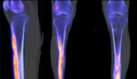
Lumbar computed tomography excluded significant nerve root compression, and venous Doppler ultrasonography ruled out deep vein thrombosis. Because of persistent symptoms and severe refractory hyperparathyroidism, whole-body bone scintigraphy with SPECT/CT was performed. Planar imaging demonstrated multiple areas of markedly increased tracer uptake involving both tibias and distal femurs (Figure 1A). Fused SPECT/CT images localized the tracer uptake to elongated intramedullary lesions in both tibias associated with cortical thickening (Figure 1B–C), consistent with multifocal BTs 23. The symptomatic right-sided lesion was identified, and revision parathyroidectomy was subsequently performed, resulting in marked symptomatic improvement one month later.
Figure 1.Imaging findings of multifocal brown tumors secondary to refractory hyperparathyroidism.
Discussion
Brown tumors are an uncommon manifestation of uncontrolled hyperparathyroidism in patients with chronic kidney disease. They result from excessive osteoclastic bone resorption and replacement of bone by fibrovascular tissue containing macrophages and multinucleated giant cells, which may produce well-defined, sometimes expansile osteolytic lesions with a characteristic “blown-out” appearance 1234. In this case, the clinical presentation was atypical, with progressive neuropathic pain initially suggesting a neurological or musculoskeletal disorder. The absence of significant lumbar nerve root compression or deep vein thrombosis, together with severe refractory hyperparathyroidism, raised the possibility of BTs.
Whole-body bone scintigraphy demonstrated multifocal skeletal involvement, revealing more extensive skeletal involvement than suggested by the focal symptoms. SPECT/CT further characterized the lesions by providing precise anatomical localization and demonstrating their intramedullary distribution and associated cortical thickening. The concordance between the symptomatic right lower limb and the corresponding tibial lesion provided a strong clinicoradiological correlation. This illustrates the complementary roles of whole-body scintigraphy and SPECT/CT: The former provides an overview of skeletal involvement, whereas the latter improves anatomical characterization and correlation with clinical findings 2.
The differential diagnosis of focal hypermetabolic bone lesions in patients with chronic kidney disease includes other metabolic bone disorders, benign bone lesions, and malignant skeletal involvement. The combination of severe refractory hyperparathyroidism, multifocal skeletal abnormalities, and the compatible imaging findings supported the diagnosis of multifocal BTs 23. Histological confirmation was therefore not deemed necessary given the clinical, biochemical, and imaging findings.
Management of BTs relies on controlling the underlying hyperparathyroidism. Medical treatment with calcimimetics and vitamin D analogs may be effective in selected patients, whereas parathyroidectomy should be considered in severe or refractory disease 13. In this case, persistent marked hyperparathyroidism despite previous subtotal parathyroidectomy and medical therapy led to revision parathyroidectomy. The subsequent marked improvement in symptoms supports a relationship between the skeletal lesion and the patient’s pain. Regression of the BTs and/or relief of local mechanical or neural effects may have contributed to the clinical improvement.
Conclusion
This case highlights that BTs should be considered in the differential diagnosis of dialysis patients with unexplained focal neuropathic pain and severe refractory hyperparathyroidism. Whole-body bone scintigraphy combined with SPECT/CT is particularly valuable by demonstrating the extent of skeletal involvement and providing precise anatomical localization with clinicoradiological correlation. Recognition of multifocal BTs may facilitate appropriate management of the underlying severe refractory hyperparathyroidism, in this case with revision parathyroidectomy resulting in marked symptomatic improvement.
Declarations
Authors' contributions:
Manuscript drafting: TP, AC (they contributed equally to this work and share first authorship); manuscript review and editing: EG, FG, ID; imaging analysis and review: OG; final approval: all authors approved the final version of the manuscript.
Informed consent:
Written informed consent was obtained from the patient.
Ethical considerations:
This study was conducted in accordance with the principles of the Declaration of Helsinki. Given the retrospective nature of the study and the use of anonymized data, formal approval by an ethics committee was not required in accordance with institutional policy.
Funding:
This research received no external funding.
Conflict of interest:
None of the authors has any conflicts of interest to declare.
Data availability:
This report is based on the patient’s medical records held by the institution. Local legislation does not permit the disclosure of patient medical records.
Use of Artificial Intelligence
Artificial intelligence tools were used for English revision and translation assistance. AI was not used for writing the manuscript itself.
ORCID IDs:
AC: https://orcid.org/0009-0007-8855-4217
TP: https://orcid.org/0009-0001-6013-5762
OG: https://orcid.org/0000-0001-8478-9675
ID: https://orcid.org/0000-0002-2108-5280
EG: https://orcid.org/0000-0001-9242-7148
GF: https://orcid.org/ 0009-0006-7087-6798
Copyright & License
© 2026 Thibault Preumont, Alexandra Corceovei , Ines Dufour, guillaume Fernandes, Olivier Gheysens, Eric Goffin.
This work is licensed under a Creative Commons Attribution 4.0 International License.

References
1. KDIGO CKD-MBD Work Group. KDIGO 2017 clinical practice guideline update for the diagnosis, evaluation, prevention and treatment of chronic kidney disease–mineral and bone disorder (CKD-MBD). Kidney Int Suppl (2011). 2017 Jul;7(1):1-59. doi: https://doi.org/10.1016/j.kisu.2017.04.001.
2. Xie C, Tsakok M, Taylor N, Partington K. Imaging of brown tumours: a pictorial review. Insights Imaging. 2019 Jul 29;10(1):75. doi: https://doi.org/10.1186/s13244-019-0757-z.
3. Santoso D, Thaha M, Empitu MA, Kadariswantiningsih IN, Suryantoro SD, Haryati MR, et al. Brown tumour in chronic kidney disease: revisiting an old disease with a new perspective. Cancers (Basel). 2023 Aug 15;15(16):4107. doi: https://doi.org/10.3390/cancers15164107.
4. Gaied H, Trabelsi M, Hadded S, Thabti Y, Khadhar M, Goucha R. Early-Onset Craniofacial Brown Tumor Progressing to Multifocal Skeletal Lesions in Peritoneal Dialysis Patient: A Case Report and Literature Review. Bull Dial Domic [Internet]. 2026 Jun. 15 [cited 2026 Sep. 6];9(2):83-92. Available from doi: https://doi.org/10.25796/bdd.v9i2.87111.
Downloads
Submitted
Accepted
Published
How to Cite
Issue
Section
License
Copyright (c) 2026 Thibault Preumont, Alexandra Corceovei , Ines Dufour, guillaume Fernandes, Olivier Gheysens, Eric Goffin
This work is licensed under a Creative Commons Attribution 4.0 International License.


